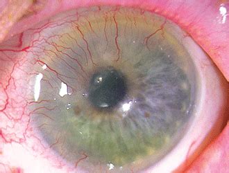
Corneal Neovascularization

Corneal neovascularization is a complex and significant ocular condition that affects the cornea, the transparent front part of the eye. It involves the abnormal growth of blood vessels into the cornea, which is typically avascular. This process can lead to various complications and impact visual acuity, making it a crucial topic of interest in ophthalmology and eye health.
Understanding Corneal Neovascularization

The cornea, a highly specialized tissue, is designed to be clear and devoid of blood vessels to maintain optimal visual function. However, certain conditions or injuries can disrupt this balance, leading to corneal neovascularization. This condition is characterized by the growth of new blood vessels from the limbal vascular plexus, located at the periphery of the cornea, into the normally avascular stromal layer.
Causes and Risk Factors
Corneal neovascularization can arise from a range of factors, including:
- Inflammatory conditions: Chronic inflammation, such as in keratitis or uveitis, can trigger the release of vascular endothelial growth factor (VEGF), promoting new blood vessel formation.
- Trauma: Physical injuries to the eye, such as corneal lacerations or burns, can initiate a healing response that includes neovascularization.
- Chemical injuries: Exposure to certain chemicals or toxic substances can damage the cornea and stimulate blood vessel growth.
- Contact lens-related issues: Prolonged or improper use of contact lenses can lead to hypoxia (oxygen deficiency) and inflammation, both of which are risk factors for neovascularization.
- Infections: Bacterial, viral, or fungal corneal infections can induce an inflammatory response that promotes angiogenesis.
- Systemic diseases: Conditions like diabetes or rheumatoid arthritis, which are associated with chronic inflammation, can increase the risk of corneal neovascularization.
Clinical Presentation
The clinical signs of corneal neovascularization can vary, but commonly include:
- Redness and irritation of the eye.
- Blurred or distorted vision.
- Sensitivity to light (photophobia).
- Visible blood vessels on the corneal surface.
- Discomfort or pain, especially with advanced cases.
Diagnosis and Assessment

Diagnosing corneal neovascularization typically involves a comprehensive eye examination. Ophthalmologists may use various techniques to evaluate the extent and severity of the condition, including:
Slit-Lamp Biomicroscopy
This is a standard method for examining the anterior segment of the eye. It allows for detailed visualization of the cornea, including the depth and pattern of neovascularization.
Corneal Staining
Dye staining, such as with fluorescein or lissamine green, can highlight areas of corneal damage or inflammation, providing valuable information about the extent of neovascularization.
Imaging Techniques
Advanced imaging technologies like confocal microscopy and optical coherence tomography (OCT) can offer high-resolution images of the corneal structure, aiding in the precise assessment of neovascularization.
| Imaging Technique | Description |
|---|---|
| Confocal Microscopy | Provides cellular-level imaging of the cornea, allowing for detailed analysis of blood vessel structure and density. |
| OCT | Uses light waves to create cross-sectional images of the cornea, aiding in the detection and measurement of neovascularization depth. |

Treatment Options
The management of corneal neovascularization aims to control the growth of new blood vessels, reduce inflammation, and preserve visual function. Treatment strategies may include:
Topical Medications
Various eye drops are used to manage corneal neovascularization. These include:
- Corticosteroids: These powerful anti-inflammatory agents can suppress the inflammatory response and slow down neovascularization. However, prolonged use can lead to complications, so they are often used short-term or in combination with other therapies.
- Non-steroidal anti-inflammatory drugs (NSAIDs): NSAIDs can help reduce inflammation and discomfort associated with neovascularization. They are often used as an adjunctive therapy.
- Anti-VEGF agents: These medications target vascular endothelial growth factor, a key driver of angiogenesis. They are particularly effective in managing neovascularization related to inflammatory conditions.
Surgical Interventions
In advanced cases or when medical management is insufficient, surgical options may be considered. These include:
- Photorefractive keratectomy (PRK): This laser procedure can be used to reshape the cornea and remove abnormal blood vessels. It is often performed in combination with other treatments.
- Corneal transplantation: In severe cases of corneal scarring or opacity due to neovascularization, a corneal transplant may be necessary to restore vision.
- Amniotic membrane transplantation: Amniotic membrane grafts can be used to promote healing and reduce inflammation in cases of corneal damage or ulceration associated with neovascularization.
Therapeutic Contact Lenses
Specialized contact lenses, such as bandage lenses or scleral lenses, can provide comfort and protection to the corneal surface, aiding in the healing process and reducing the need for frequent medication administration.
Prognosis and Long-Term Management
The prognosis for corneal neovascularization varies depending on the underlying cause and the extent of vascularization. Early diagnosis and prompt treatment can lead to better outcomes and potentially prevent permanent vision loss.
Long-Term Management Strategies
For patients with corneal neovascularization, ongoing care is essential to monitor the condition and manage any associated complications. This may include regular eye examinations, continued use of prescribed medications, and adherence to contact lens care protocols.
Prevention and Risk Reduction
Preventive measures are crucial in reducing the risk of corneal neovascularization. This includes proper eye protection to prevent injuries, careful contact lens care and hygiene, and timely management of inflammatory or infectious eye conditions.
Research and Future Directions

Ongoing research in corneal neovascularization aims to improve our understanding of the condition and develop more effective treatments. Current areas of focus include:
Advanced Imaging Technologies
Researchers are exploring the use of cutting-edge imaging techniques to provide more detailed and precise assessments of corneal neovascularization. This includes the development of artificial intelligence-based algorithms for automated analysis of corneal images.
Novel Therapeutic Approaches
New medications and therapeutic strategies are being investigated to target the underlying mechanisms of neovascularization. This includes the development of sustained-release drug delivery systems and gene therapy approaches to modulate angiogenic factors.
Clinical Trials and Collaborative Research
Numerous clinical trials are underway to evaluate the safety and efficacy of emerging treatments for corneal neovascularization. Collaborative efforts among ophthalmologists, researchers, and industry partners are essential to advance our knowledge and improve patient outcomes.
Conclusion
Corneal neovascularization is a complex ocular condition that requires careful diagnosis, management, and ongoing care. With early intervention and appropriate treatment, many patients can experience improved visual outcomes and maintain their quality of life. As research continues to advance, we can expect to see more effective and targeted therapies, ultimately improving the prognosis for those affected by this condition.
What are the potential complications of corneal neovascularization?
+Corneal neovascularization can lead to various complications, including reduced visual acuity due to scattering of light by the new blood vessels, increased risk of infection, and potential for permanent scarring of the cornea. In severe cases, it can result in corneal edema, bullous keratopathy, or even corneal perforation.
Can corneal neovascularization be prevented?
+While it may not always be preventable, certain measures can reduce the risk. These include proper eye protection to prevent injuries, good contact lens hygiene and care, and prompt management of inflammatory or infectious eye conditions. Maintaining overall eye health and regular eye examinations are also crucial for early detection and intervention.
How effective are anti-VEGF medications in treating corneal neovascularization?
+Anti-VEGF medications have shown promising results in managing corneal neovascularization, particularly in cases associated with inflammation. These medications target vascular endothelial growth factor, a key driver of angiogenesis. However, the effectiveness may vary depending on the severity and underlying cause of neovascularization, and they are often used in combination with other treatments.


